Case 46: Complete surgical phase: Maxillary arch 4 implants and Mandibular arch- Guided Bone regeneration with Titanium mesh and Autogenous bone graft and collagen membrane
Case 46: Complete surgical phase: Maxillary arch 4 implants and Mandibular arch- Guided Bone regeneration with Titanium mesh and Autogenous bone graft and collagen membrane
Here is a case which is affected by generalised periodontitis in both maxillary and mandibular arch along with failed implants in the lower anteriors with severe bone defects.
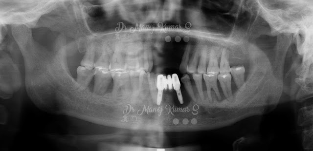


Here is a case which is affected by generalised periodontitis in both maxillary and mandibular arch along with failed implants in the lower anteriors with severe bone defects.

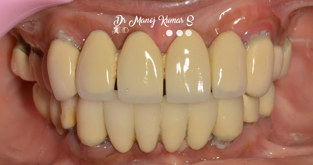
All the teeth were mobile and the plan was to go in for a total extraction followed by a wait period of 2-3 weeks before going ahead with implants in the maxillary arch ( 4 numbers) and a bar supported hybrid prosthesis with shortened arch. In the lower arch Guided bone regeneration at the lower anterior defect site extending from 4.3 to 3.4. Since the plan is to place implants in the sites of A to E (According to Misch) and a lower hybrid prosthesis with shortened arch over 5 implants.
The first phase or the surgical phase was handed over to me to place the implants and the guided bone regeneration procedure.
After a span of 2-3 weeks we opened the maxillary site for implants. Prior CBCT assessment made us plan 4 implants in the site of 13 and 15 in first quadrant and 24 and 26 in the second quadrant. A complete flap was raised to visualise the bone from molar to molar region and a palatal holding suture was place for easier visualisation and flap handling. The bone was irregular and was flattened using an osteotomy bur in a straight hand piece. Straumann implants RC were placed at the desired sites.


After a week the lower arch guided bone regeneration was carried out. A full flap was raised from 3.7 to 4.7 and the defect site was curetted before grafting as the site was a previous failed site of implants. Then Autogenous bone was collected from 4.7 and 3.7 sites and a total of 0.5 cc collected from the Bonesier drill. This autogenous bone was mixed with 0.25 cc of Bio-oss xenograft to attain more bulk.


Later the defect was packed after creating multiple bleeding sites created on the site on the surface of the boneusing a small round bur, where the graft is intended to be placed to fulfill its blood supply. The graft is packed and then a Titanium mesh35x 45 mm was used and adapted on the labial aspect and tac screws were placed to hold the mesh. the mesh was wrapped around and held again on the lingual side by a set of tacs. Further collagen membrane was placed over the mesh before achieving a complete closure of the soft tissue.


The lower arch is to be planned for implant after 6 months. In-between there is a possibility of the mesh to get exposed. If that happens anytime sooner than 6 weeks then it is ideal to wait till 12 weeks before removing them. Until then hygiene measures are to be employed by the patient.
this cas ewas done by me at Parasu Dental Hospital, Chennai.

Comments