Posts

Showing posts from January, 2020

Case 4: Severely discolored stump- Zirconia As the savior

Image
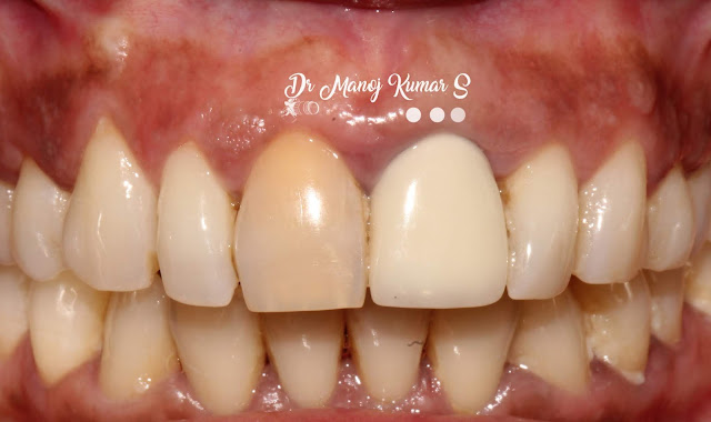
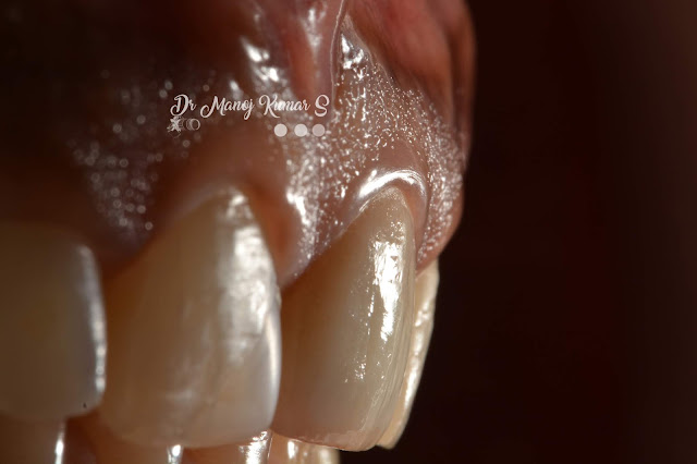
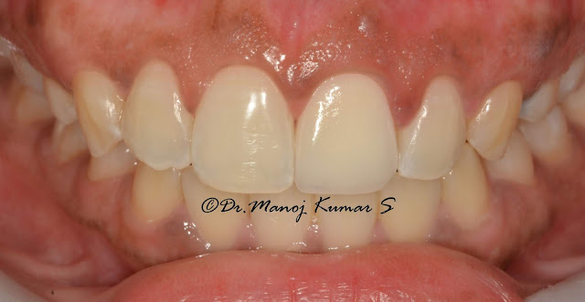
Case 4: Severely discolored stump- Zirconia As the savior Not always emax is going to be suitable. Certain situations like the one we are going to have a look, where the underlying dentin is terribly discolored needs a masking effect in the crown. This patient came with dis-satisfied anterior metal ceramic crown along with a discolored non vital maxillary central incisor. Initially, I had thoughts of doing an internal bleach in 1.1 and remove the crown in 2.1 and assess. But after removal of crown in 2.1 I had doubts in bleaching the underlying discolored stump. It was blackish and i decided to choose Zirconia with an opaque core as more recent zirconias have improved tranlucency. This eventually made me go ahead with two crowns rather than doing just a single crown. I prefer cementing Zirconia crowns with GIC cement and that has been a personal choice, although there is a huge debate in choosing a resin cement with necessary surface treatment on the intaglio surface of zir

Case 3: Emax single crown in tooth 2.1

Image
Case 3: Emax single crown in tooth 2.1 Here is another case on managing a severely discolored non-vital endodontically treated tooth. we did give the option of bleaching followed by the choice on need for a crown. Owing to time restraint the patient opted for a quicker esthetic solution and went ahead with the option of restoring the esthetics with an E-max crown in 2.1 The situation involved a minor midline diastema. A choice of veneer on 11 and a crown in 21 to close the space was also opted out and the patient was contempt with just an esthetic restoration in 2.1 The sequence of treatment involved indexing for a luxatemp provisional, followed by crown preparation and elastomeric impression followed by shade mapping and provisionalisation. The luting cement that was used in this case was Variolink -translucent resin cement. The crown material was an Emax- Cad milled on an MT blank. This case was done completely by me at Parasu Dental Hospital, Chennai.

Publication 2: Marginal fit and microleakage of cast and metal laser sintered copings—An in vitro study

Purpose This study aimed to compare the marginal fit and microleakage of metal laser sintered Co–Cr alloy copings and conventional cast Ni–Cr alloy copings using a stereomicroscope. Methods Forty extracted maxillary  premolars  were randomly divided into two groups. One group was subjected to coping fabrication using conventional lost wax (LW) technique while the other group was subjected to coping fabrication using metal laser sintering (MLS) technology. The marginal fit of these copings were compared before and after ceramic addition using images obtained with a steromicroscope and an ImageJ analysis software. All the specimens were cemented using Type 1  glass ionomer cement  and were subjected to thermocycling. The specimens were evaluated for microleakage using stereomicroscope and 2%  methylene blue  die used as a  tracer . The data were subjected to statistical analysis using paired  t -test,  Mann–Whitney test  and  Chi-Square test . Results The mean marginal f

Case 2: E-max Crown in 2.1- Esthetics mimicked

Image
Case 2: E-max crown in 2.1 All ceramic restorations are considered the best in achieving the esthetics. e-max crowns are indeed 'Esthetic Maximum' crowns. Here is a case of discolored non vital 2.1 tooth which needed esthetic enhancement. Considering the crown length discrepancy and tooth being endodontically treated, I suggested for an E-max crown for the best esthetic result. Shown below is the pre-op image of the clinical situation. The subsequent steps involving crown preparation, provisionalisation with Luxatemp provisionals followed by cementation and esthetic dental photography can be seen as follows. The health of the gingiva and the location of finishline and the emergence from the soft-tissue are few aspects which need attention for an ideal mimicking crown restoration. Definitive cementation was done with Variolink Resin cement - Translucent shade. This case was done by me at Parasu Dental Hospital Chennai

Case 1: Achieving the desired emergence profile - IMPLANT ESTHETICS

Image
Case 1: Immediate loading Dental Implant in 2.2 and Soft tissue Esthetics Achieving the desired emergence profile in anterior dental implants are always challenging. Here is a case of a single immediate loaded implant in tooth 2.2 which was done with Nobel active narrow implants. Immediate loading was done with chair side composite on temporary screw retained abutment. One can see how the soft tissue gets molded over a span of 5 months and the gingival cuff that is achieved is commendable.  The method of using a stock impression coping and customising the emergence profile with the use of existing provisional crown as an index, is one of the easiest method in analog dentistry. Ofcourse, digitial intra oral scanners are available which makes dentists’ job simple. However, here you can see the sequential set of images with description within on how the emergence can be created and replicated in your impression. The type of prosthesis which was decided was a custom zirconia abutme

Publication 1: Virtual Smile Design- A contemporary field in Digital Dentistry

Image
Creating templates for smile designing with Photoshop software. Computer design software has become a primary tool for communication among the dentist, patient, and ceramist. Virtual smile design can be carried out using various software programs, most of which use assorted forms of teeth templates that are made based on the concept of "golden proportion." Despite current advances in 3-dimensional imaging and smile designing, many clinicians still employ conventional design methods and analog (ie, man-made) mock-ups in assessing and establishing esthetic makeovers. To simplify virtual smile designing, the teeth templates should be readily available. No literature has provided details as to how to create these templates. This article explains a technique for creating different forms of teeth templates using Adobe Photoshop® CS6 that eventually can be used for smile design purposes, either in Photoshop or Microsoft Powerpoint. Clinically speaking, various smile design